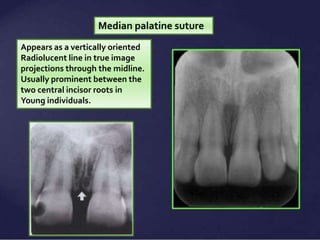
incisive foramen radiograph
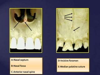
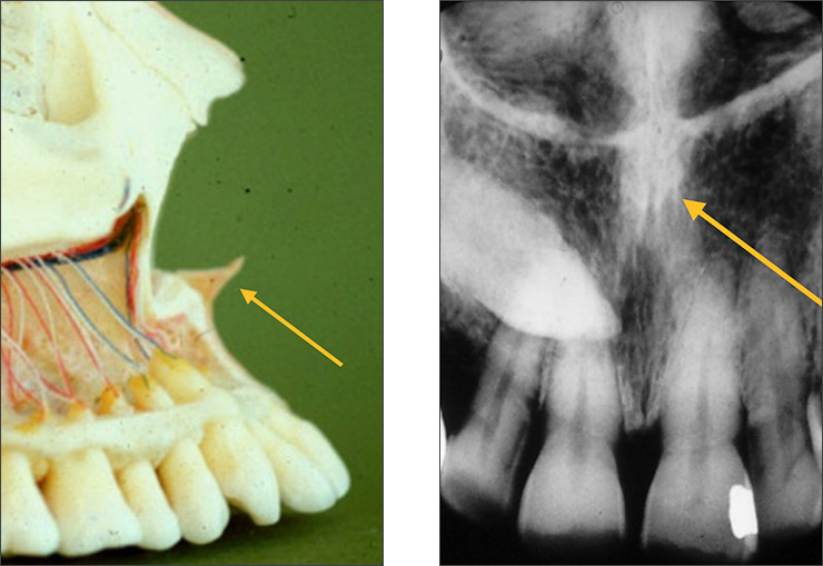
The incisive foramen is situated within the incisive fossa of the maxilla. Transmit nasopalatine nerves and branches of the descending palatine artery.

Maxillary Anterior Landmarks Intraoral Radiographic Anatomy Continuing Education Course Dentalcare Com
Coronoid process is the thin triangular-shaped process of the anterosuperior aspect of the ramus.

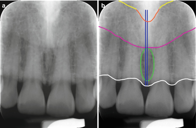
. Incisive foramen seen as an ovoid radiolucent area in the midline of the maxilla between the roots of the central incisors. Yellowfloor of nasal cavity. It is located in the maxilla in the incisive fossa midline in the palate posterior to the central incisors at the junction of the medial palatine and incisive sutures.
Or nasopalatine foramen is a round to oval radiolucent structure located in between the roots of the maxillary central incisors. The canal of each side courses forward obliquely converging toward the nasal septum and then descends in a vertical direction to pass through the Y-shaped incisor foramen or anterior palatine canal in the hard palate. Reported that the diameter of the nasal and oral openings was higher in men compared to women.
It will appear as a round to ovoid radiolucent area between the roots. Exit through Foramina of Stenson. The incisive foramen also known as nasopalatine foramen or anterior palatine foramen is the oral opening of the nasopalatine canal.
Opening or hole in bone that permits the passage of nerves and blood vessels b. It is opens between the roots of the maxillary central incisors on the lingual. Anterior palatine foramen or nasopalatine foramen is the opening of the incisive canals on the hard palate immediately behind the incisor teethIt gives passage blood vessels and nerves.
The radiolucency results from a depression above and posterior to the lateral incisor. Assessments included 1 mesiodistal diameter 2 labiopalatal diameter 3 length of the incisive canal 4 shape of incisive canal and 5 width of the bone anterior to the incisive foramen. The median suture of the palate see figure 3-23 may appear as a radiolucent line extending posteriorly from the alveolar border.
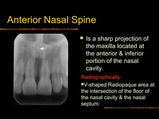
The following characteristics of incisive were evaluated. Incisive foramen 9 Anterior nasal spine Landmarks in the Maxilla a floor of nasal fossa b maxillary sinus c lateral fossa d soft tissue of the nose Maxillary Canine d c b a Lateral fossa. Tap card to see definition.
Lateral canals on each side of the midline. The diameter of the incisive foramen is on average less than 6 mm in the dentulous maxilla. Orange V-shaped lineanterior nasal spine.
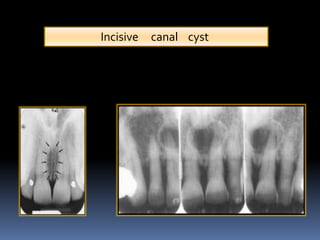
Root resorption and tooth displacement may be present. Pinksoft tissues of the nose. It can be single or multiple.
On a mandibular periapical radiograph the mental foramen appears as a small ovoid or round radiolucent area located in the apical region of the mandibular premolars. A The incisive foramen also called nasopalatine oranterior palatine foramen Fig. Article in Croatian Cvetković T.
Approximately 30 of cases contain respiratory epithelium 3. In general inter-root distance between medial points of central incisors roots and incisive canal opening was greater in the region close to upper central incisors apex mean 384 mm. What is the nasopalatine incisive foramen Click card to see definition.
They may appear heart-shaped if the anterior nasal spine is superimposed. Broad shallow scooped-out or depressed area of bone c. It is a V shaped radiopaque area situated at the intersection of.
Radiographic features They are seen as a solitary well-defined oval or round unilocular radiolucency between central incisors 6 mm in diameter. It is actually in the anterior part of the palate but superimposition makes it appear to be located between the roots of the central incisors. 1 Width of the nasopalatine canal labiopalatally and mesiodistally Figures 1 a and 1 b 2 Length of the canal Figure 1 c 3 Width of the bone anterior to the canal Figure 1 d 4 Shape of the canal Figures 2 a 2 d.
Failure to protect the mental foramen may lead to permanent loss of normal sensation in the lower lip. 2A 3A is seen as an oval radiolucency between the roots of the maxillary central inci- sors. In radiographs exposed from the region of the cuspid or lateral incisor the incisive foramen may appear as a radiolucency at the apex of one of the incisors.
Interpretation of incisive foramen on radiographs. SD 144. Hollow space cavity or recess in bone d.
The mean length of the incisive canal was. The incisive foramen is the inferior opening of the nasopalatine canal incisive canal. The incisive foramen is used as an.
Panoramic radiography can help locate the mental foramen through which the nerve supply to the lower lip passes and the mandibular canal during dental implantations 14. It is an opening or hole in bone located on the external surface of the mandible in the region of the mandibular premolars. Sharp thornlike projection of bone Opening or hole in bone that permits the passage of nerves and blood vessels.
A b Maxillary incisors periapical in a dentate patient. Mean canal length was 1863 235 mm and males. Parallel blue linesintermaxillary suture median palatal suture.
The mean width of the foramen labiopalatally and mesiodistally was 312 094 mm and 323 098 mm respectively. Its appearance is quite variable due to normal anatomic variation and due to the operators angulation of the x-ray beam. On periapical x-ray images the incisive foramen is located in the midline between the roots of the central incisors.
Whitealveolar ridge Full size image Fig. There were 9 parameters measured regarding to incisive foramen including the incisive canal length and buccal bone width millimeters mmResults. In the human mouth the incisive foramen also known as.
The canal terminates in the roof of. This measurement is consistent across ethnicities while some also report minor differences between the sexes. It also appears that the diameter increases.
Sinus canal A foramen is a n.

Opg Showing Incisive Foramen And Mental Foramen Download Scientific Diagram
An Example Of A Large Incisive Canal Mesial To The Mental Foramen The Download Scientific Diagram
Cone Beam Computed Tomography Assessment Of The Maxillary Incisive Canal And Foramen Considerations Of Anatomical Variations When Placing Immediate Implants Plos One

Incisive Foramen Dr G S Toothpix
Normal Radiographic Anatomical Landmarks

Mouth Incisive Canal Cyst Professional Radiology Outcomes

Figure 2 Assessment Of The Mandibular Incisive Canal By Panoramic Radiograph And Cone Beam Computed Tomography

Normal Radiographic Anatomical Landmarks

Visibility Of Mandibular Anatomical Landmarks In Panoramic Radiography A Retrospective Study Semantic Scholar
Normal Anatomical Landmarks In Dental X Rays And Cbct Springerlink

Pdf The Evaluation Of Visibility Of Mandibular Anatomic Landmarks Using Panoramic Radiography Semantic Scholar

A Panoramic Radiograph Shows The Anterior Loop And Incisive Canal Download Scientific Diagram
Normal Radiographic Anatomical Landmarks
Intra Oral Radiographic Anatomical Landmarks
Maxillary Anterior Landmarks Intraoral Radiographic Anatomy Dentalcare
Normal Radiographic Anatomical Landmarks

Periapical Radiograph 1 Year After Treatment Bone And Teeth Showing Download Scientific Diagram
2

6 Essentials Of Dental Radiographic Analysis And Interpretation Pocket Dentistry


Comments
Post a Comment